
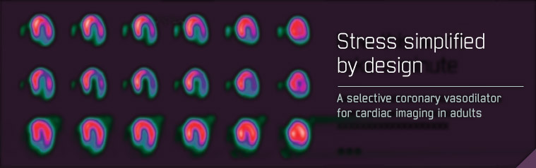
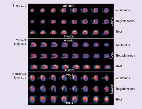
Patient image example
SPECT perfusion images after adenosine and regadenoson stress and at rest in the same patient performed on different days according to the ADVANCE MPI protocol. A lateral perfusion defect is noted on both stress images.1
Figure: Reproduced with permission1
References
- Zoghbi G and Iskandrian A. Regadenoson Myocardial Perfusion Imaging. Imaging Med 2010; 2:395-406.